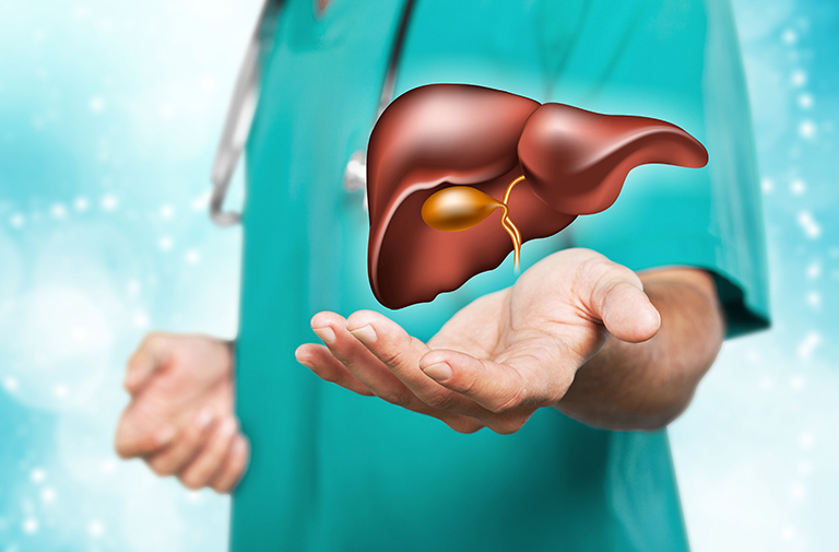
Bile Ducts
BILE DUCTS
Like small branches of a tree, there are many small bile ducts inside the liver (intrahepatic). These form the right and left bile ducts at the lower edge of the liver and outside (extrahepatic). The right and left bile ducts unite to form the main hepatic bile duct, and with the opening of the cystic duct coming from the gallbladder to this duct, the common bile duct (choledoch) is formed. The common bile duct opens into the duodenum and drains the bile into the intestine. The place where the choledoch opens into the intestine is called the oddi canal, and the most distal point of this canal is called the ampulla. In 90% of patients, the pancreatic duct opens to the last part of the common duct and forms a common duct and pours into the duodenum. In 10% of patients, they are open into the intestine separately.
The liver makes bile and the bile ducts carry the bile made by the liver and the contents of the gallbladder to the intestine. Bile is necessary for the digestion of dietary fats from the intestines.
When there is an obstruction (stone, tumor, benign stricture, etc.) anywhere in the bile duct, the bile cannot be fully discharged into the intestine, the bile ducts expand, bile accumulates in the blood and jaundice develops.
BILE DUCT CANCER (CHOLANGIOCARCINOMA)
Cancer arising from the cells (epithelium) of the biliary tract. It can originate from any part of the biliary tree. It is called intrahepatic cholangiocarcinoma if it originates from the biliary tract in the liver tissue, and extrahepatic cholangiocarcinoma if it originates from the biliary tract outside of the liver tissue. If extra-hepatic cholangiocarcinoma arises from the junction of the right and left biliary tract, it is called Perihilar biliary tract cancer (Klatskin tumor).
About 20% of all cholangiocellular cancers are of intrahepatic origin. Biliary intraepithelial neoplasia (BillN) and intraductal papillary neoplasia (IPNB) of the biliary tract are considered to be cholangiocarcinoma precursor lesions.
WHAT ARE THE RISK FACTORS THAT CAUSE THE CHOLANGIOCARCINOMA?
There are several established risk factors for developing cholangiocarcinoma;
- Primary sclerosing cholangitis (PSC); an inflammatory disease of the biliary tract (cholangitis)
- Choledochal cyst; congenital cystic dilatation of the biliary tree.
- Caroli's disease; congenital disorder characterized by multifocal, segmental dilatation of large intrahepatic bile ducts.
- Ulcerative colitis; inflammatory disease of the bowel often accompany PSC
- Hepatolithiasis (stones in the hepatic biliary tract)
- Anastomoses between the bile duct and the intestine
- Bile duct infections and some parasitic diseases
- Cirrhosis
- Hepatitis B or Hepatitis C virus
- Advanced age
- Obesity; by increasing the formation of stones in the biliary tract or by causing some hormonal changes
- Family history; having a family history of bile duct cancer
- Some chemicals; Exposure to dioxin, nitrosamine, polychlorinated biphenyl, which can be seen more especially in plastic and automotive industry workers.
- Diabetes
- Alcohol
- Other possible risk factors; smoking, pancreatitis (inflammation of the pancreas), HIV infection
WHAT ARE THE SYMPTOMS SEEN IN CHOLANGIOCARCINOMA?
- Jaundice
- Itching
- Weight loss
- Anorexia
- Fever
- Abdominal pain
WHAT ARE THE SYMPTOMS SEEN IN CHOLANGIOCARCINOMA?
- Taking the patient's history and physical examination
- Tumor markers, CEA (carcinoembryonic antigen), CA 19-9; They are found above normal values in blood, urine and tissues. They can also be elevated in some other types of cancer. There is no rule that they will always be high in a patient with cancer, sometimes they can be found in normal values.
- Biopsy; It is taking a piece of tissue thought to be cancerous and examining it under a microscope. However, this may not always be possible.The biopsy can be taken during ERCP, PTK, or it can be taken percutaneously under the guidance of radiology with a needle inserted directly into the cancer tissue.
- Imaging methods;
- Ultrasonography
- Computed tomography
- Magnetic resonance imaging
- PTC (percutaneous transhepatic cholangiography); Under the guidance of radiology, a needle is inserted through the skin into the biliary tract in the liver, and a X-ray film is taken by injecting a contrast agent into the biliary tract. Both the bile ducts are displayed and if there is an obstruction or narrowing in the biliary tract, this is revealed. At the same time, bile can be taken out of the body by means of a tube (stent) left in the biliary tract, or the narrow area can be expanded to allow the bile to flow into the intestine. Thus, the jaundice of the patient, if any, is eliminated.
- ERCP (endoscopic retrograde cholangiopancreatography); A finger-thick flexible tube with a video camera system at the end is inserted through the mouth, passing through the esophagus and stomach, and reaching the hole where the bile duct opens into the duodenum. If there is a tumor a biopsy can be taken and if the tumor causes a stenosis, a tube (stent) left in the bile duct that can be used to bypass the narrow area and allow the bile to flow into the intestine.
- Endoscopic Ultrasonography (EUS); a finger-thick flexible tube with a video camera system and ultrasonography probe at the end is inserted through the mouth and passed through the esophagus and stomach to reach the hole where the bile duct opens into the duodenum. With the ultrasonography performed in this area, the presence of tumor and its extent to the surrounding tissues, vessels and lymph nodes are investigated, if necessary, a biopsy is taken.
- Laparoscopy; It is the visual evaluation of the gallbladder, biliary tract, liver and other organs by entering the abdomen with a thin tube with a video camera and light source at the end. It helps us to obtain information about the stage of the cancer and, if necessary, to take a biopsy. It gives an idea whether the cancerous tissues can be completely removed.
- Cholangioscopy; It can be done during ERCP. With a very thin fiber optic tube and camera, it allows us to directly enter the biliary tract and see the cause of obstruction (stone, tumor, etc.) and, if necessary, take a biopsy.
STAGING IN CHOLANGIOCARCINOMA
- Local; the cancer is limited to the biliary tract and can be surgically removed. There is no spread of cancer beyond the biliary tract.
- Locally advanced; the cancer also affected the biliary tract and adjacent organs and vessels. There is no evidence that the cancer has spread to distant parts of the body (metastasis).
- Metastatic; the cancer has spread to distant parts of the body.
TREATMENT IN CHOLANGIOCARCINOMA
While determining the treatment plan, it is decided by looking at the location and extent of the cancer, whether it can be surgically removed or not, some side effects of the treatment, whether it will prolong the patient's life span and whether it will positively affect the quality of life.
- Surgery
- emoval of the biliary tract; If the cancer is limited to the biliary tract, the biliary tract and regional lymph nodes are removed.
Removal of some liver tissue along with the bile duct; if the cancer is close to the liver or in the intrahepatic biliary tract
Whipple procedure; If the cancer is close to the pancreas, in the area where the bile duct opens into the duodenum (cancer of the ampulla), whipple operation is performed. The biliary tract, gallbladder, head of the pancreas, part of the stomach, part of the small intestine are surgically removed.
Liver transplantation; Since the probability of recurrence is very high, this treatment can only be applied in a carefully selected group of patients. - Radiotherapy (Radiation Therapy)
- High-energy X-rays are aimed at destroying cancer cells.
- It can be used in the treatment of the disease, as well as to control the symptoms and pain in advanced disease.
- Fatigue, mild skin reactions, nausea, diarrhea can be seen as side effects.
- Chemotherapy
- It is the use of drugs to damage cancer cells and prevent their growth and proliferation.
- Chemotherapy is used to reduce tumor size before surgery or in patients who cannot undergo surgery. For this purpose, intravenous or oral medications can be used.
- Palliative Treatments
- Treatments to reduce and eliminate these side effects are called palliative treatments. For example, relief of pain, strengthening of nutrition and giving additional nutritional foods.
- Stent placement and surgical bypass procedure; Sometimes a metallic or plastic stent is placed in the blocked biliary tract in patients where the cancer has spread too far to be removed completely, and a new pathway is opened between the bile duct above the obstruction and the small intestine below the obstruction. In this approach, it is aimed to increase the patient's comfort of life, it is not possible to completely remove the cancerous tissues.